Gliomas are the most common intracranial primary malignant tumors, accounting for approximately 80% of all malignant brain tumors, and are characterized by high recurrence rates, high mortality rates, and low cure rates. Surgery is the preferred option for glioma treatment. However, due to the infiltrative growth characteristics of gliomas in the brain, it is often difficult for surgeons to accurately define the boundaries of gliomas for guiding their resection intraoperatively, resulting in a high recurrence rate of gliomas after surgery. Glioma localization techniques such as magnetic resonance and intraoperative fluorescence currently used in clinical practice are mainly based on tumor-induced destruction of vascular structures, which outline the area of blood-brain barrier disruption but do not completely overlap with the area of glioma infiltration. Accurate localization of glioma-infiltrating areas remains a challenge due to the decreasing density of cancer cells at the glioma margins, which diminishes the specificity and sensitivity of cancer-targeted imaging techniques. Therefore, the development of new strategies for intraoperative identification of glioma infiltrating regions is important for improving surgical prognosis. Recently, Professor Cong Li's group at the School of Pharmacy, Fudan University, proposed a new strategy to locate glioma infiltrative margins by visualizing immunosuppressive macrophages. The related work was published online in Advanced Science under the title "Intra-operative Definition of Glioma Infiltrative Margins by Visualizing Immunosuppressive Tumor Associated Macrophages".

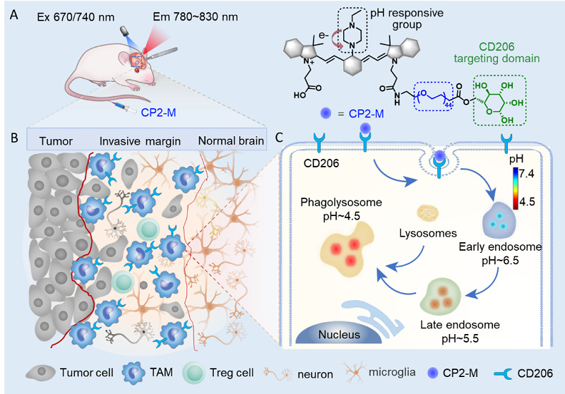
Visualization of immunosuppressive tumor-associated macrophages localizing glioma boundaries. The probe CP2-M was first targeted to the CD206 receptor overexpressed by immunosuppressive macrophages through a hierarchical targeting strategy, and then activated in the acidic microenvironment of phagolysosomes after CD206 receptor-mediated endocytosis to achieve accurate localization of immunosuppressive macrophages.
By analyzing glioma databases and tumor specimens from glioma patients, the content of M2-TAMs in gliomas was significantly higher than that in normal tissues, and immunohistochemical staining revealed the aggregation of M2-TAMs at the glioma invasion margins, which led to the scientific hypothesis that M2-TAMs serve as a new target for localizing glioma invasion boundaries. This strategy defines the glioma invasion margin by visualizing M2-TAMs rather than cancer cells and is therefore not affected by the decreasing density of cancer cells at the tumor margin. To achieve precise localization of M2-TAMs in vivo, a hierarchical targeting strategy was proposed in this study. The pH-ratio responsive fluorescent probe CP2-M was first targeted to the CD206 receptor on the immunosuppressive macrophage, and after CD206 receptor-mediated endocytosis the probe was activated in the acidic microenvironment of macrophage phagocytic vesicles to achieve accurate localization of immunosuppressive macrophages. In vivo imaging studies based on both rat and nude mouse glioma models demonstrated that CP2-M was able to specifically target brain gliomas and locate intracranial glioma infiltration with a high signal-to-noise ratio. In addition, CP2-M outperformed the clinically used glioma navigation probes 5-ALA and indocyanine green (ICG) in prolonging post-operative survival. The results of behavioral experiments showed that model rats did not cause significant neurological impairment after CP2-M-guided surgery. The above studies suggest that the immunosuppressive TAMs-targeted strategy provides a new pathway to overcome the navigation distortion caused by the reduced density of cancer cells at the glioma border, improve the surgical resection rate of gliomas, reduce the postoperative recurrence rate, and prolong the survival of patients.
Ph.D. candidate Chong Cao and Hang Yin at the School of Pharmacy, Fudan University, attending neurosurgeon Qi Yue and Ph.D. candidate Biao Yang at Huashan Hospital are the co-first authors of the paper. Professor Ying Mao of Huashan Hospital affiliated to Fudan University, Researcher Xiaoyong Zhang from Institute of Science and Technology for Brain-Inspired Intelligence, Fudan University, Young Researcher Xiao Zhu and Zuhai Lei, Professor Cong Li at School of Pharmacy, Fudan University are the corresponding authors of the paper. The research was also supported by Professor Jinhua Yu from the School of Information Science and Technology, Fudan University. This research work was funded by the National Natural Science Foundation of China and the Science and Technology Support Project of Shanghai Science and Technology Commission in the field of biomedicine.
Original link: https://onlinelibrary.wiley.com/doi/10.1002/advs.202304020