Surgeons face challenges in intraoperatively defining margin of brain tumors due to its infiltrative nature. Extracellular acidosis caused by metabolic reprogramming of cancer cells is a reliable marker for tumor infiltrative regions. There is a spatiotemporal correlation between tissue acidity and malignancy in solid tumors. Moreover, acidity promotes tumor progression by establishing an immunosuppressive environment and driving glioma cells to a pluripotent, stem cell-like state by upregulating stem cell markers. Intraoperatively visualizing and excising the acidic regions holds the promise to improve surgical prognosis.
Recently, Professor Cong Li (School of pharmacy, Fudan University), Professor Xiaoyong Zhang (Institute of Science and Technology for Brain-Inspired Intelligence, Fudan University), Professor Jinhua Yu (School of Information Science and Technology, Fudan University) and Professor Ying Mao (Huashan Hospital) jointly built a glioma surgery navigation system based on surface enhanced Raman scattering (SERS) technology.
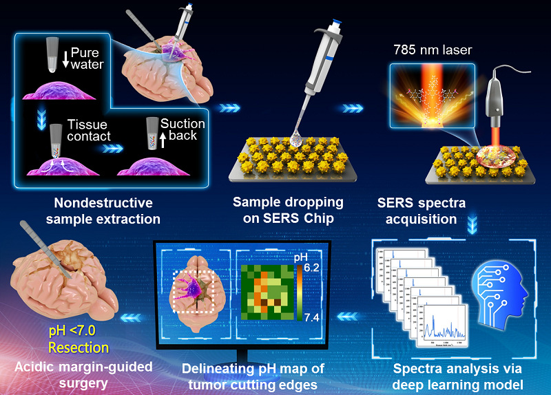
This system is composed of a SERS chip that senses sample’s acidity via pH-responsive Raman signals, a handheld Raman scanner that collects the Raman signal of the sample placed on the SERS chips, and a homemade deep learning model that determines sample’s pH by automatically processing the Raman spectra. The intelligent SERS navigation system can measure the pH of 64 tissue sites (about 1 cm2) within 6 minutes, and delineate the metabolically acidic margin by drawing the pH map. Acidity correlated cancer cell density and proliferation level were demonstrated in tumor cutting edges of animal models and excised tissues from glioma patients. The overall survival of animal models post the SERS system guided surgery was significantly increased in comparison to the conventional strategy used in clinical practice. This SERS system holds the promise in accelerating clinical transition of acidic margin-guided surgery for solid tumors with infiltrative nature.

Figure 1. Schematic diagram of the SERS navigation system intraoperatively delineating acidic margin of glioma. A trace amount of pure water (≈ 0.4 μL) in the pipette tip contacts suspicious tissue at the tumor cutting edge for 2‒4 s. Then, the water droplet is sucked back and dripped onto a homemade pH-sensitive SERS chip. The Raman spectra of the aqueous sample on the SERS chip were acquired by a handheld Raman scanner equipped with a 785 nm laser. The pH map of tumor cutting edges was intra-operatively delineated with the assistance of a deep learning model by automatically analyzing the Raman spectra. With the guidance of the pH map, acidic tissues with pH values less than 7.0 were excised.
The surgical navigation system can quickly locate the glioma infiltration area during operation and improve the surgical prognosis of glioma animal model. In addition, This SERS system holds the promise in accelerating clinical transition of acidic margin-guided surgery for avoiding inject exogenous probes.
The work was published online in Advanced Science under the title of Intelligent SERS navigation system guiding brain tumor surgery by internally defining the metallic acidosis. Ziyi Jin (PhD student, school of pharmacy), Qi Yue (attending physician, Huashan Hospital), Wenjia Duan (PhD student, school of pharmacy) and An Sui (master student, Department of Electronic Engineering), share the co-first authorship of the paper. Professor Ying Mao (Huashan Hospital), Professor Jinhua Yu (Fudan University), Professor Xiaoyong Zhang (Fudan University) and Professor Cong Li (Fudan University) are the co-corresponding authors.
This work was supported by National Key Research & Development Project, the Ministry of Science and Technology of China, the Program of Shanghai Science and Technology Committee, National Science Fund for Distinguished Young Scholars, National Natural Science Foundation of China, Shanghai Municipal Science and Technology Major Project and Shanghai Center for Brain Science and Brain-Inspired Technology, “Double First-Class” initiative of Fudan University.
Introduction to Professor Cong Li
Cong Li, Professor, School of Pharmacy, Fudan University, Shanghai, China. Director of Center of Radiopharmacy and Molecular Imaging, School of pharmacy, Fudan University, PI of Key Laboratory of Intelligent Drug Delivery, Ministry of Education. He was funded by “National Science Fund for Distinguished Young Scholars, China”, “National Key Research & Development Project, China”, etc. He was awarded Second Class Prizes of The State Scientific and Technological Progress Award (2015), New Century Excellent Talents of Ministry of Education of China (2013), Young Scientific Talents of Shanghai (2016) and “Dawn Scholar” Program of Shanghai Education Commission (2015). His research work focuses on the visualization and regulation of immune microenvironment of brain diseases, radioactive diagnosis and treatment drugs, and the research and development of intelligent surgical navigation equipment. Professor Li has published many academic papers in Nat Biomed Eng, Adv Mater, Angew Chem Int Ed, Adv Sci and other journals, and obtained 6 patents. He currently holds positions of Deputy Secretary General, Biological Photonics Committee, Chinese Society of Biomedical Engineering; Committee Member, Brain Tumor Committee, Chinese Medical Doctor Association; and Committee Member, Molecular Imaging Committee, Chinese Society of Biophysics.
