由于独有的多通道分辨能力,超高的灵敏度和超低的检测限,表面增强拉曼散射技术(Surface Enhanced Resonance Scattering, SERS)已在生物医学领域显示出巨大的应用价值。手持式拉曼成像仪也已成功应用于临床肿瘤诊断、恶性分级和手术导航。然而皮肤,黏膜等表面组织强光散射系数严重削弱拉曼成像仪对活体组织的成像深度和成像质量,阻碍了该技术大规模临床转化。
5月5日在线发表于《ACS Applied Materials & Interfaces》杂志上的一项研究中“Noninvasively Imaging Subcutaneous Tumor Xenograft by a Handheld Raman Detector with the Assistance of an Optical Clearing Agent”,复旦大学药学院李聪教授的研究团队报道了光透明技术能够显著性降低活体组织皮肤散射,有效改善SERS成像灵敏度从而实现手持式拉曼成像仪对皮下肿瘤模型的高信噪比示踪。

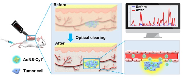
图1. 光透明技术实现皮下肿瘤的高信噪比SERS成像
该论文第一作者张云飞同学介绍:通过制备基于金纳米星的超灵敏度(5 fM)SERS纳米探针,联合光透明技术降低表皮组织的散射系数,实现了对皮下肿瘤的高信噪比无创示踪。通讯作者李聪教授介绍:该研究的创新点在于首次将光透明技术引入SERS活体肿瘤成像领域。通过降低活体皮肤组织的散射系数并结合超灵敏的SERS探针,使手持式拉曼成像仪观察到更深,更微小的病灶,拓展SERS成像技术的临床应用范围。

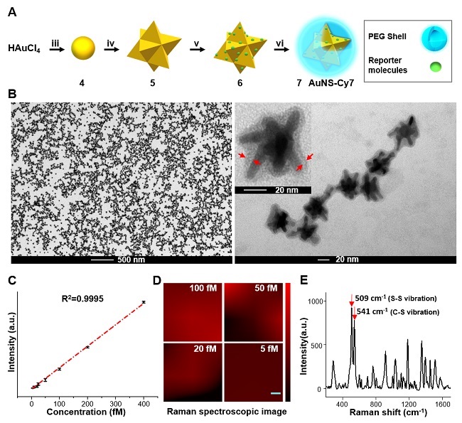
图2. 超灵敏SERS探针的合成和表征。(A)拉曼探针合成示意图;(B)拉曼探针TEM示意图;(C?D)探针信号强度随浓度变化示意图;(E)拉曼探针特征光谱图。

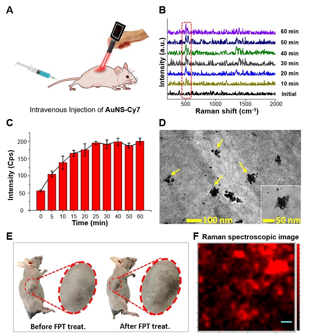
图3. 手持式拉曼成像仪无创观察皮下瘤模型。(A)无创肿瘤SERS成像示意图;(B)施加光透剂后肿瘤区域随时间变化的SERS光谱;(C)施加光透剂后肿瘤区域随时间变化的SERS信号强度;(D)TEM图揭示探针肿瘤内存在;(E)肿瘤区域施加光透剂前后白光示意图;(F)肿瘤切片拉曼显微镜成像。
Title: Noninvasively Imaging Subcutaneous Tumor Xenograft by a Handheld Raman Detector with the Assistance of an Optical Clearing Agent
DOI: 10.1021/acsami.7b04205
http://pubs.acs.org/doi/abs/10.1021/acsami.7b04205
Abstract: A handheld Raman detector with operational convenience, high portability, and rapid acquisition rate has been applied in clinics for diagnostic purposes. However, the inherent weakness of Raman scattering and strong scattering of the turbid tissue restricts its utilization to superficial locations. To extend the applications of a handheld Raman detector to deep tissues, a gold nanostar-based surface-enhanced Raman scattering (SERS) nanoprobe with robust colloidal stability, a fingerprint-like spectrum, and extremely high sensitivity (5.0 fM) was developed. With the assistance of FPT, a multicomponent optical clearing agent (OCA) efficiently suppressing light scattering from the turbid dermal tissues, the handheld Raman detector noninvasively visualized the subcutaneous tumor xenograft with a high target-to-background ratio after intravenous injection of the gold nanostar-based SERS nanoprobe. To the best of our knowledge, this work is the first example to introduce the optical clearing technique in assisting SERS imaging in vivo. The combination of optical clearing technology and SERS is a promising strategy for the extension of the clinical applications of the handheld Raman detector from superficial tissues to subcutaneous or even deeper lesions that are usually “concealed” by the turbid dermal tissue.


